In other words: methylation patterns in meningiomas – originating from the meninges – are very different from the methylation seen in sarcomas, which begin in connective tissues.
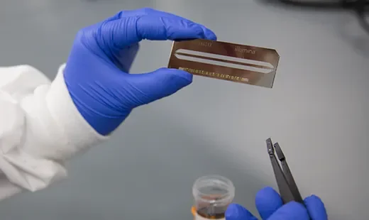
The JAX APML began offering methylation profiling to outside institutions in 2022 and has processed about two hundred tumor samples each year since. When a patient with brain cancer either undergoes a biopsy or has surgery to remove a tumor, a cluster of cancer cells are isolated and sent to the APML.
“JAX is really well-suited to be able to do this,” says Kelly. “We already had the equipment and expertise that not many labs in the country have.”
Qian Wu, a neuropathologist at UConn Health, is among those who collaborated with JAX to launch methylation profiling. She now sends about a quarter of all the new brain tumors samples she encounters to the APML for testing.
“With brain tumors, getting a biopsy at all is incredibly difficult and we sometimes only get a tiny amount of tissue that is really hard to work with,” explains Wu. “In the past, we put those cells under a microscope and tried to make a diagnosis based on how they looked. Methylation profiling gives us a lot more data with our limited samples and acts as an independent way of diagnosing brain tumors.”
Wu explains that DNA methylation analysis is considered the best method of diagnosing brain tumor severity. While she can identify a tumor’s type under a microscope, grading its aggressiveness (on a scale of 1 to 4) is much harder. Methylation analysis helps refine this grading process.