
A closer look at endometriosis pain: New map reveals key cellular interactions
By Roberto Molar Candanosa
Article | October 9, 2026
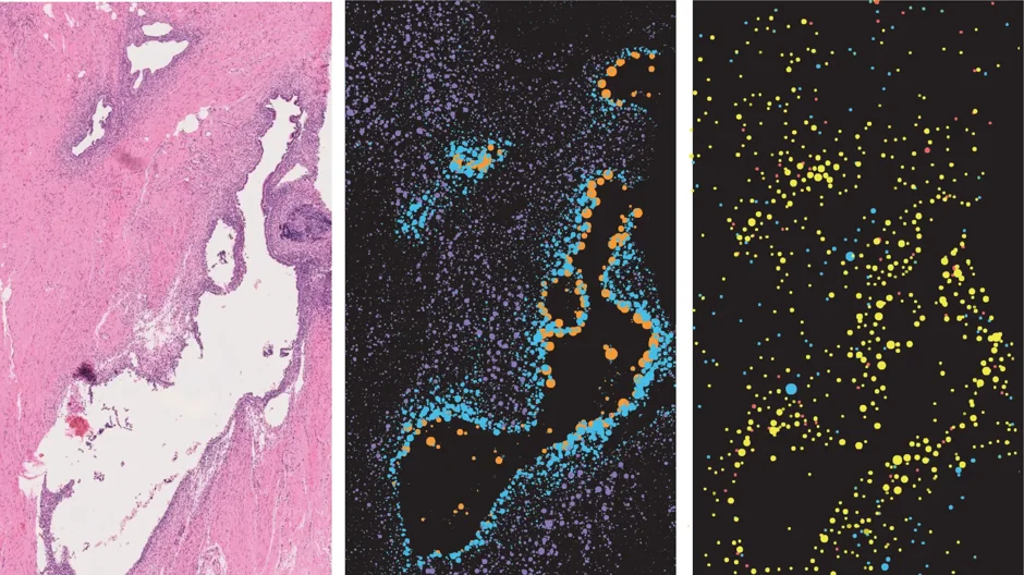
People with endometriosis often report struggling for years to have their agonizing pelvic pain, severe period cramps, and other life-impacting symptoms taken seriously by doctors. New research from The Jackson Laboratory (JAX) may help change that, as scientists have mapped the endometriosis lesion microenvironment with unprecedented detail.
Published in Nature Communications, the study explains how the interplay between neurons and immune cells amplifies pain signaling. It’s the first time the team has used human tissue samples from the EndoRISE biorepository in Connecticut, the only state-funded program in the United States that pairs comprehensive endometriosis sample collection and distribution with education, advocacy, and awareness.
“Doctors typically minimize your symptoms, saying menstrual cycles are normally painful and that others feel the same pain,” said Caroline Haney, a JAX developmental biologist and endometriosis patient who led the research. “It turns out that’s not the whole story. The pain is not just in your head; it’s a biological reaction, actual pain signaling coming from the neurons in your body.”
Endometriosis is a chronic, systemic disease that affects about 10% of female-born individuals. It occurs when tissue similar to the lining of the uterus (called endometrium) grows outside the uterus, forming lesions. This can lead to inflammation, scarring, debilitating pain, and infertility in some cases. Why it spreads, how it causes pain, and what drives the disease remain poorly understood, making diagnosis and treatment a persistent challenge.
Pioneering science
Directed by JAX’s Elise Courtois, Assistant Professor and Director of the Single Cell Biology Lab, the team surveyed samples from the two most common endometriosis lesion areas, the ovaries and the peritoneum, a membrane lining the abdominal and pelvic cavity. They mapped stromal cells, which provide structural and functional support to organs, and epithelial cells using advanced spatial technologies that provide a single cell-resolution map of the lesion spatial architecture.
Traditional technology often can’t easily map neurons. To pinpoint these cells, the researchers used high‑resolution spatial transcriptomics. This revealed which genes are active and exactly where cells sit relative to one another inside intact tissue. The result was sharp maps of tissue structure, nerve connections, and cell groupings near endometriosis lesions.
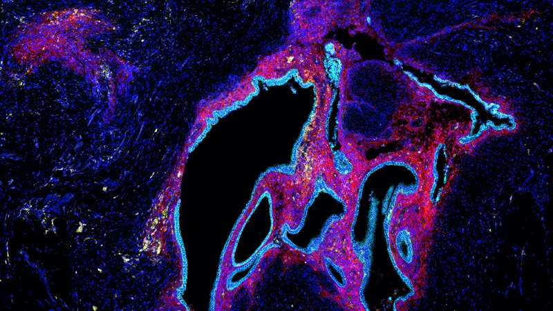
The new maps revealed that lesions cells organize in distinct layers around epithelial glands, a key feature of endometriosis lesions. Areas closest to the glands, called “close stroma” were enriched in MME+ fibroblasts, activated immune cells such as macrophages, and pain‑sensing nerve cells. In “far stroma,” different fibroblasts (OGN+) and immune cells like B cells and mast cells were more common. This pattern was consistent across samples in both ovarian and peritoneal lesions.
“We typically think of ovarian and peritoneal as two different lesion types—and they do have differences—but it seems their underlying cellular architectures are very similar,” Haney said.
The data also showed that nociceptive neurons, which transmit pain, cluster near epithelial glands, where they sit close to activated macrophages and certain fibroblasts that may help promote nerve growth. These patterns point to a neuro-immune feedback loop that drives chronic pain and inflammation, with the strongest signaling occurring in the close stroma next to epithelial glands. There, MME+ fibroblasts, activated macrophages, and nociceptor neurons interact to amplify pain signaling, promote inflammation, and remodel tissue.
Farther from the glands, these interactions are reduced, and nociceptive signaling drops off sharply. Other sensory neuron subtypes in the far stroma, including mechanoreceptors and proprioceptor-like neurons, are more broadly distributed, with mechanoreceptors more often found alongside different immune cells. This offers a new picture of how different neuron subtypes are organized within these lesion environments.
Haney, who recently underwent laparoscopic surgery to have her endometriosis lesions removed, said she is confident the data will help researchers find more effective treatments. Currently, diagnosis can take years, and the only definitive answer comes from surgery. Treatments include over‑the‑counter pain relievers, hormonal therapy such as birth control, and surgeries that don’t always end symptoms or prevent lesion recurrence and disease progression.
“Current treatment options for endometriosis do not work for everyone, and when they do work, the relief is sometimes only temporary. Our research, which uncovers the neurons involved in lesion derived pain, will help the field find better options for pain management,” Haney said.
Recreating pain with organoids
To confirm their observations, the researchers also grew peripheral sensory nerve organoids, three-dimensional tissue models made from human induced pluripotent stem cell (iPSC)-derived cell lines generated at JAX. By combining the neuron organoids with human epithelial cells and fibroblasts grown from patient-derived lesions, the researchers confirmed that epithelial cells paired with fibroblasts attracted neurons toward a pain-sensing, inflammatory state.
A 3D lab model shows pain‑sensing neurons extending from a human stem‑cell‑derived nerve organoid (center) toward cells taken from endometriosis lesions (surrounding clusters). The model provides evidence that endometriosis lesions actively promote nerve growth and pain‑related signaling, interactions also observed in patient tissue samples. Credit: Caroline Haney, Ph.D., The Jackson Laboratory.
Courtois, a molecular biologist and lead study author, said the team’s next focus will be to integrate more immune components into the complex in vitro models and evaluate different therapeutic approaches to break the neuroinflammatory process induced by lesion components.

“We’re trying to make the most of these technologies to dive deeper into the endometriosis pathobiology that we’ve known about for more than a hundred years,” said Courtois, who also co-directs EndoRISE. “The EndoRISE biorepository is a key part of this effort, giving us access to the samples we need to perform this research and ultimately move toward a definitive, more effective cure.”
The study was made possible by the endometriosis tissue biorepository housed at JAX's Genomic Medicine campus in Farmington, Connecticut and run by EndoRISE, a partnership program between JAX and UConn Health. EndoRISE leads the biorepository program and was instrumental in enabling this research. UConn Health and other hospital partners provide many of the samples, which come from individuals who were assigned female at birth, ages 18 and above, who consented and enrolled in the program. The study was supported by the Mayday Fund.
Other authors are Elaheh Alizadeh, Joshua Lee, Meryl Sullivan, Jasmina Kuljancic, William F. Flynn, Paul Robson, and Brian S. White of The Jackson Laboratory; and Danielle E. Luciano of University of Connecticut Health Center.
JAX media contact: Patrick Skahill, [email protected], 860-839-3309.
Learn more about The Jackson Laboratory.
Citation: Haney, C., Alizadeh, E., Lee, J. et al. High-Resolution Spatial Transcriptomics Reveals Fibroblast and Neuroimmune Microenvironments in Endometriosis Lesions. Nature Communications. (2026). DOI: 10.1038/s41467-026-78453-5
1 in 10: The endometriosis challenge
EndoRISE is more than a research initiative. It’s a model for how states can take on complex, underfunded diseases in a coordinated, impactful way.
View more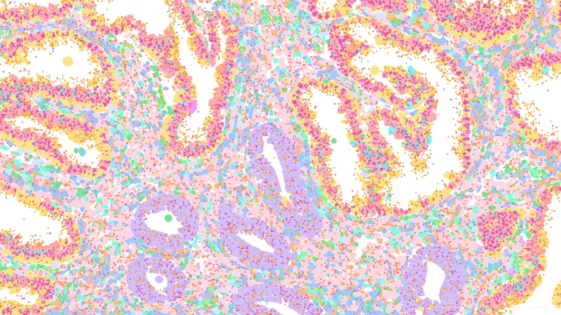
Endometriosis
Endometriosis affects 1 in 10 female-born individuals globally, causing pain and infertility. Diagnosis is challenging, often requiring surgery.
View more