Painless skin patch offers new way to monitor immune health
Article | March 2, 2026
Microneedle patch samples key immune cells often missed in routine blood draws and biopsies
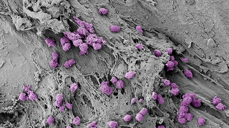
(Farmington, Conn. – March 2, 2026) – Researchers at The Jackson Laboratory (JAX), in collaboration with the Massachusetts Institute of Technology (MIT), have developed the first bandage-like microneedle patch that can sample the body’s immune responses painlessly from the skin. The device detects inflammatory signals within minutes and collects specialized immune cells within hours without the need for blood draws or surgical biopsies.
Already, the patch is helping researchers and clinicians study immune responses in aging and skin autoimmunity, including vitiligo and psoriasis. In the future, it could make it easier to track how people respond to vaccines, infections, and cancer therapies by complementing traditional blood tests and biopsies while being far easier on patients.
“Traditionally, studying some of the most important immune cells in the body requires a skin biopsy or blood draws. Because many of these cells live and respond in tissues like the skin, accessing them has meant invasive procedures,” said Sasan Jalili, a biomedical engineer and immunologist at JAX, who is also a joint faculty member at UConn School of Medicine. “We’ve shown we can capture them painlessly and noninvasively instead. This is especially important in sensitive or visible areas like the face or neck, where people often don’t want biopsies because of scarring, as well as for older adults, frail patients, and very young children or infants.”
Initially developed during Jalili’s postdoctoral training at MIT, the platform was further refined, optimized, and advanced from mouse models toward clinical application at JAX through collaborations with the University of Massachusetts Chan Medical School (UMass Chan).
Leveraging a natural immune alarm system
Most tests for monitoring immune cells and inflammatory biomarkers rely on bloodwork, but many of the cells that recognize specific infections, vaccines, or autoimmune triggers circulate only sparsely in blood.
The patch works by harnessing resident memory T cells, immune sentinels that live in skin and other “barrier” tissues and rapidly respond to previously encountered foreign threats, or antigens. When these cells recognize a familiar antigen, such as a fragment of a virus or an allergen, they “sound the alarm,” releasing signals to attract additional immune cells from the bloodstream, including the highly specialized T cells that recognize that same threat.
By triggering this natural process, which concentrates key immune cells in the skin, the researchers deliberately assessed immune responses. The sampled material revealed the number and state of T cells and other signaling molecules, offering a dynamic readout of the immune system’s strength and responsiveness to specific diseases and conditions.
In this study, we used antigen-specific T cells as a proof of concept, but the patch also captures other immune cells and inflammatory biomarkers.
Sasan Jalili
In mouse vaccination models, the patch dramatically boosted the recovery of antigen-specific T cells, recruiting many of these cells from the bloodstream rather than skin. In a human test at UMass Chan, the patch also collected a rich mix of immune cells and signaling proteins, including resident memory T cells.
“This study marks the first demonstration of live human immune cell sampling using a microneedle patch,” Jalili said. “This opens the door to a new way of monitoring immune responses that’s practical, painless, and clinically feasible.”
Expanding the immune monitoring toolbox
The patch may be especially useful for skin conditions, since immune cells that drive conditions such as allergic dermatitis, psoriasis, and vitiligo already live in the tissue. Jalili is already using it to study how age-related skin changes contribute to chronic inflammation and frailty in older adults as part of the Pepper Scholars Program in the UConn School of Medicine and UConn Center on Aging.
Animation displaying the network of polymers within the hydrogel on the microneedle skin patch
"People wouldn’t need hours of sampling. Even 15 to 30 minutes can be enough to detect inflammatory signals and get a sense of what’s happening in the tissue,” Jalili said.
Blood tests and biopsies will remain essential tools, and additional studies to determine how the patch performs across different diseases and patient populations are under way. But the early findings are particularly promising, said study co-author Darrell Irvine, an immunologist and bioengineer at Scripps Research, who began the work at MIT.
“Not only did we run extensive preclinical experiments, we were able to carry out an initial test in humans,” Irvine said. “That’s exciting because it almost never happens with brand-new technologies. Moving new technologies from the lab to testing on patients often takes years.”
The patch may be especially useful for skin conditions, since immune cells that drive conditions such as allergic dermatitis, psoriasis, and vitiligo already live in the tissue. Jalili is already using it to study how age-related skin changes contribute to chronic inflammation and frailty in older adults as part of the Pepper Scholars Program in the School of Medicine at UConn Health.
Looking ahead, the patch could eventually support at-home monitoring, allowing patients with skin conditions to track unpredictable flare-ups. The technology could also be adapted for oral or nasal cavities, opening the door to monitoring mucosal immune responses.
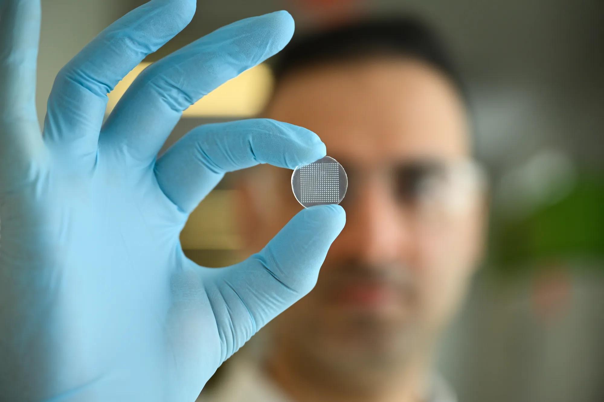
Sasan Jalili holds the microneedle skin patch which is about the size of a quarter.
Other authors in this study are Ryan R. Hosn of Massachusetts Institute of Technology; Wei-Che Ko and Khashayar Afshari of University of Massachusetts Chan Medical School; Ashok Kumar Dhinakaran of The Jackson Laboratory; Namit Chaudhary and Laura Maiorino of Massachusetts Institute of Technology; Nazgol Haddadi of University of Massachusetts Chan Medical School; Anusha Nathan, Matthew A. Getz, and Gaurav D. Gaiha of The Ragon Institute of Massachusetts General Hospital; Mehdi Rashighi and John E. Harris of University of Massachusetts Chan Medical School; and Paula T. Hammond of Massachusetts Institute of Technology, and the Koch Institute for Integrative Cancer Research.
This work was supported by the NIH (award U01AI176310), The Jackson Laboratory, the Ragon Institute of MGH, MIT and Harvard, and the Koch Institute Support Grant P30-CA14051 from the National Cancer Institute.
The authors have submitted a patent application filed by MIT related to the data presented in this work.
The Jackson Laboratory (JAX) is an independent, nonprofit biomedical research institution with a National Cancer Institute-designated Cancer Center. JAX leverages a unique combination of research, education, and resources to achieve its bold mission: to discover precise genomic solutions for disease and empower the global biomedical community in the shared quest to improve human health. Established in Bar Harbor, Maine in 1929, JAX is a global organization with nearly 3,000 employees worldwide and campuses and facilities in Maine, Connecticut, California, Florida, New York, and Japan. For more information, please visit www.jax.org.
Learn more
The Jalili Lab at The Jackson Laboratory
The Jalili Research Group at The Jackson Laboratory (JAX) is focused on interrogating the host immunity, microbiome and their interface using different engineering tools in the context of infectious diseases, autoimmunity and cancer.
JAX in Motion seeks to inspire and move audiences through a series of short documentaries. Presenting the groundbreaking research and discoveries being made by the exceptional scientific researchers at The Jackson Laboratory.